Technology

Precision Meets Innovation.

Modern diagnostic and surgical technology helps specialists detect conditions earlier, measure more precisely and plan treatment with confidence. Here is what each one does — in plain language.

Configurable sample equipment list
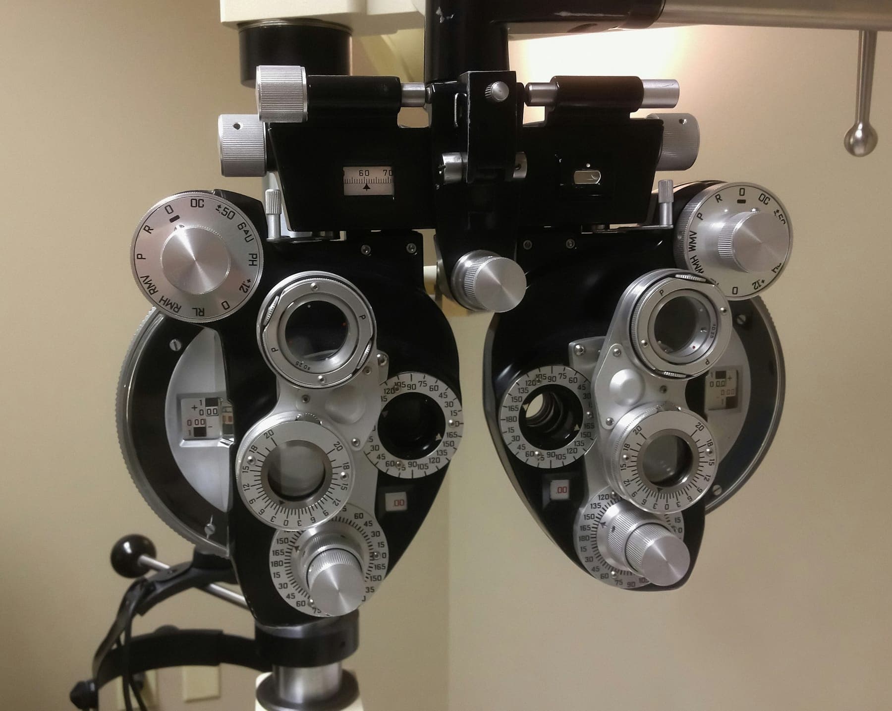
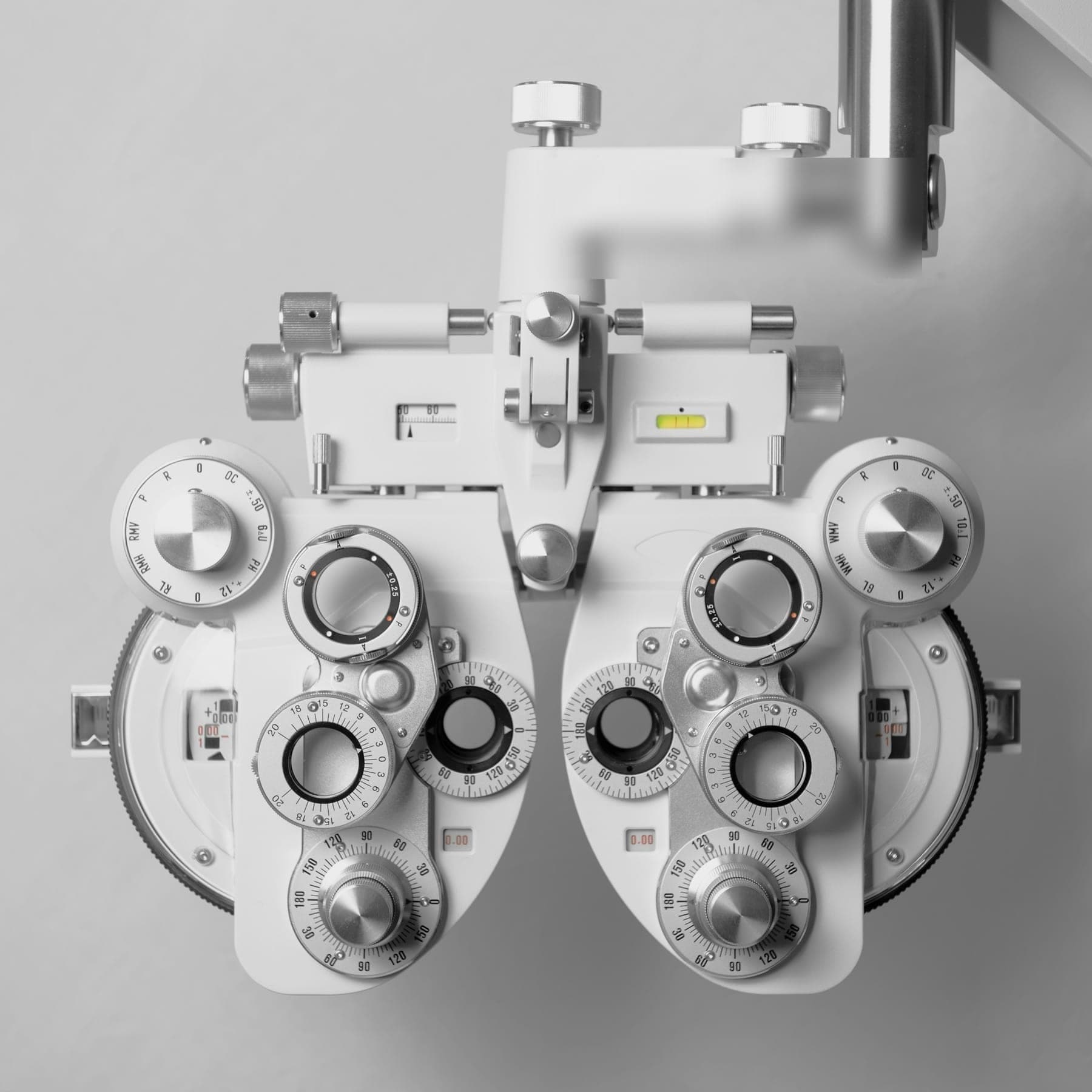
White ophthalmic phoropter instrument

Diagnostic equipment

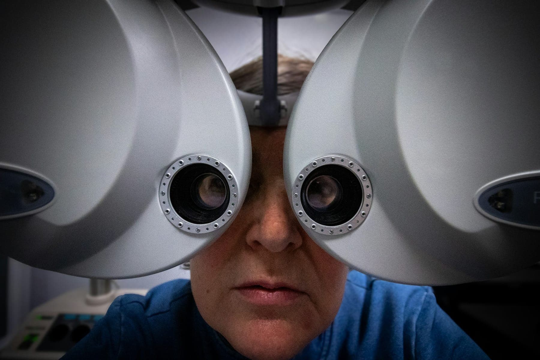
Patient looking through a phoropter during measurementDiagnostic

Biometry

Optical Biometry

Precise eye measurements used to calculate intraocular lens power for cataract surgery.

Optical biometry measures the length of the eye, the curvature of the cornea and the depth of the front chamber using light-based interferometry. These measurements feed lens-power formulas that help surgeons select an intraocular lens suited to each eye.

  • Supports accurate lens selection
  • Quick, contact-free measurement
  • Improves surgical planning
Close view of an ophthalmic refraction instrumentDiagnostic

Topography

Corneal Topography

Colour-coded maps of corneal shape for keratoconus screening and refractive planning.

Corneal topography and tomography map the curvature and thickness of the cornea. They are essential for detecting keratoconus, fitting specialist contact lenses and assessing suitability for laser vision correction.

  • Early detection of corneal irregularity
  • Essential for LASIK screening
  • Guides contact lens fitting
Clinician adjusting a trial frame on a patient's faceDiagnostic

Visual Fields

Automated Visual Field Testing

Maps your peripheral and central vision to detect functional loss.

During a visual field test you look at a central target and press a button when you notice small lights appearing around it. The result maps the sensitivity of your field of vision, which is key in diagnosing and monitoring glaucoma and neuro-ophthalmic conditions.

  • Detects loss patients may not notice
  • Tracks glaucoma progression
  • Helps localise neurological causes

Imaging systems

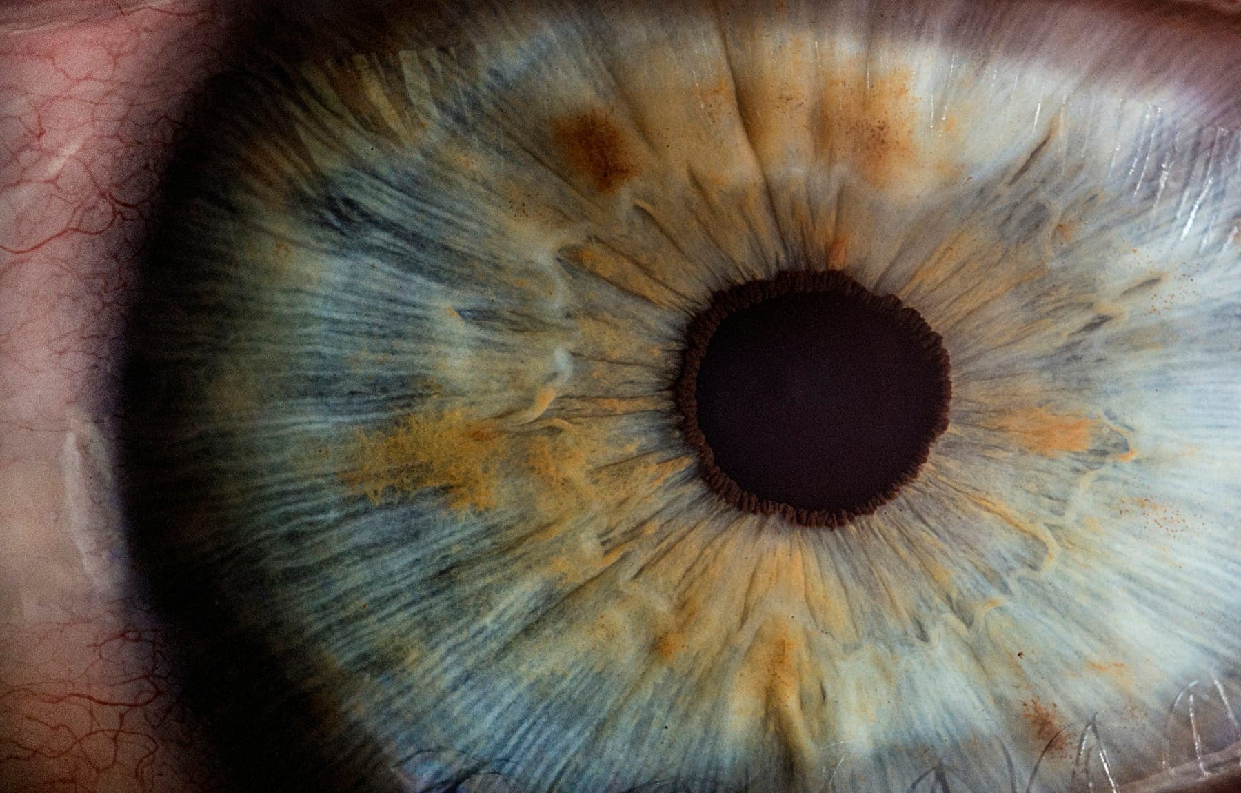
Macro photograph of a human iris and pupilImaging

OCT

Optical Coherence Tomography

Cross-sectional scans of the retina and optic nerve, captured in seconds without contact.

OCT uses near-infrared light to create high-resolution, cross-sectional images of the retina's layers, much like an ultrasound uses sound. It helps detect and monitor macular degeneration, diabetic macular oedema, macular holes and glaucoma-related nerve fibre thinning.

  • Non-invasive and contact-free
  • Micrometre-level detail of retinal layers
  • Ideal for tracking change over time
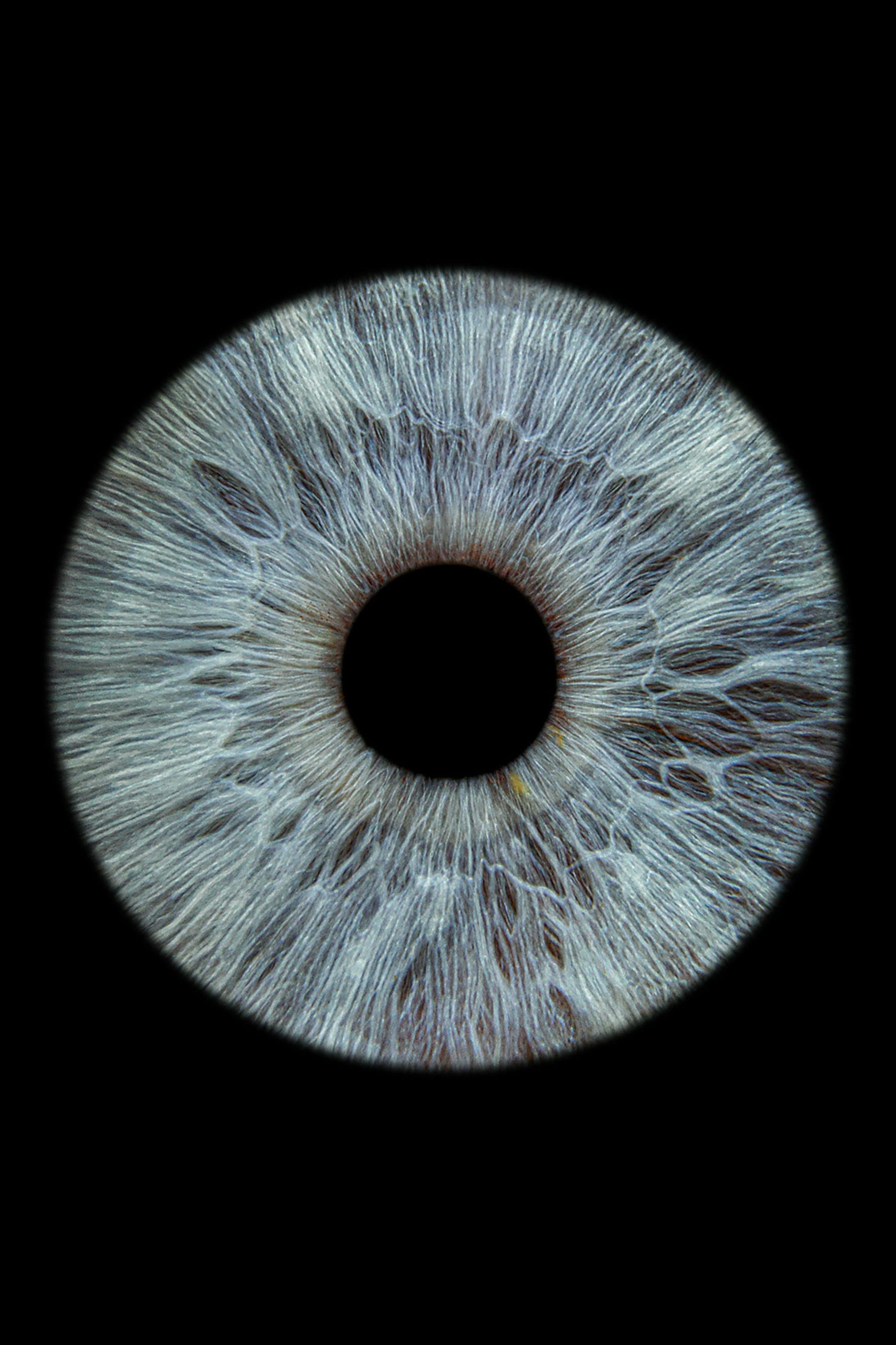
Detailed photograph of a grey-blue iris on a black backgroundImaging

Fundus Imaging

Fundus Photography

Detailed colour photographs of the retina, optic disc and blood vessels.

A specialised camera photographs the back of the eye, creating a permanent record of the retina, macula and optic nerve. Images can be compared between visits to identify subtle changes in conditions such as diabetic retinopathy and glaucoma.

  • Permanent visual record
  • Easy comparison over time
  • Helpful for patient explanation

Surgical technologies

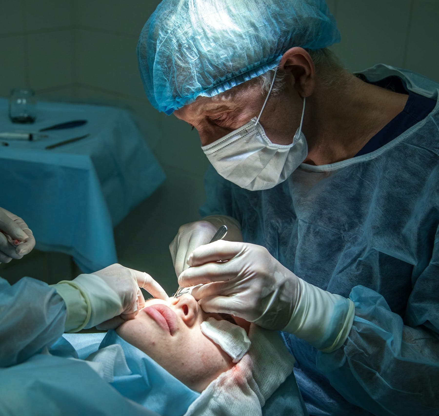
Surgeon in a cap and mask operating on a patient's eyeSurgical

Phaco

Advanced Phacoemulsification Systems

Small-incision cataract removal using controlled ultrasound energy.

Phacoemulsification systems use ultrasound to break up the clouded lens so it can be removed through a very small incision, before a folded intraocular lens is inserted. Modern fluidics help maintain a stable eye during surgery.

  • Small, often stitch-free incisions
  • Typically a day procedure
  • Supports faster visual recovery
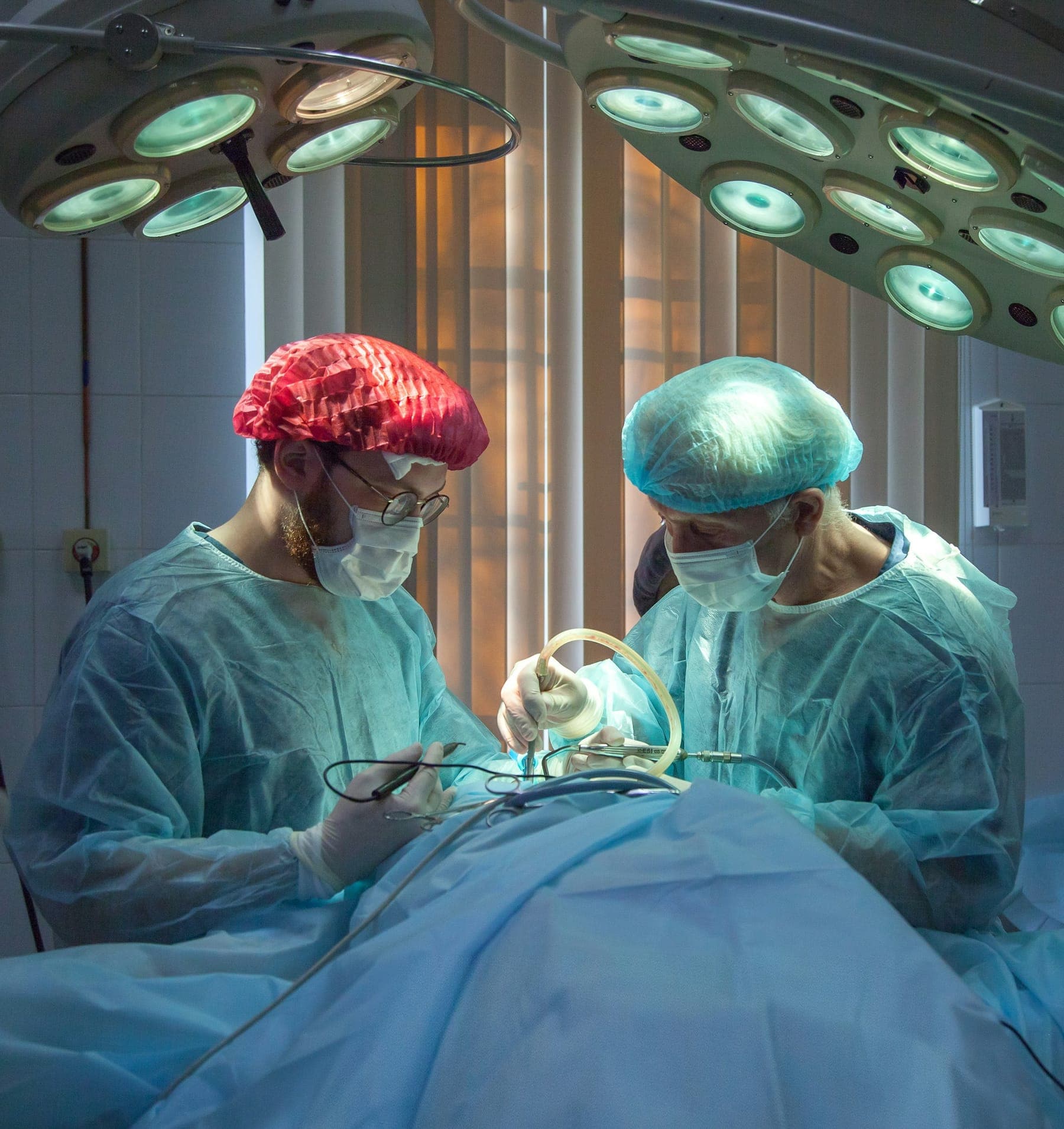
Two surgeons in scrubs operating under bright theatre lightsSurgical

Excimer Laser

Excimer Laser Technology

Ultraviolet laser that reshapes the cornea to correct refractive error.

Excimer lasers remove microscopic amounts of corneal tissue with high precision to reshape its curvature, correcting myopia, hyperopia and astigmatism in LASIK and surface laser procedures. Eye-tracking systems follow small eye movements during treatment.

  • Sub-micron precision
  • Eye tracking during treatment
  • Used in LASIK and PRK

Patient benefits

What modern technology means for you.

  • Earlier detection

    High-resolution imaging can reveal subtle changes before symptoms appear.

  • Clearer explanations

    Seeing your own scans makes conditions and decisions easier to understand.

  • Precise planning

    Accurate measurements support surgical planning and lens selection.

  • Comfortable testing

    Most diagnostic tests are quick, painless and non-contact.

FAQs

Questions about tests & scans

No. OCT, biometry, topography and fundus photography are non-contact or very gentle tests. You simply look at a target for a few seconds.

Appointments

Your Journey to Better Vision Starts Here.

Request a consultation with the right specialist. Our patient coordinators will contact you to confirm a suitable time.